
Page 62 - 07
P. 62
–1
The peaks at about 3000…2750 cm were characteristic of–CH 3 bonds. The si-
milar peaks in the FTIR spectra of the treated and untreated samples showed that there
was no change in the basic structure of nanocomposite membranes. The increments in
–1 –1
the absorption bands of C–O at 1030 cm and C=O at 1770 cm were attributed to the
–1
creation of unsaturated – C=C− bonds at 1645 cm after plasma treatment. It was also
observed from FTIR spectra that C–C and C–H bands decreased after plasma treatment.
It indicated that the cross linking phenomenon enhanced during plasma treatment. The
–1
Co NPs showed its characteristic peak at 765 cm for nanocomposite membrane.
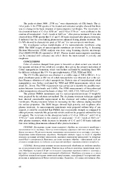
We investigated surface morphologies of Co nanocomposite membrane using
SEM. The SEM images of nanocomposite membrane are shown in Fig. 6. Scanning
Electron Microscopic (SEM) analysis was done using Scanning electron microscope
(Carl ZEISS EVOR-18) operated at 20 kV. Plasma treated nanocomposite membrane
can be compared with untreated one which shows the improvement in porosity and
roughness.
CONCLUSIONS
Color of solution changed from green to brownish as plant extract was mixed in
the aqueous solution of the cobalt ion complex, thus giving the primary indication of
Cobalt nanoparticles formation, which was further confirmed by analyzing these NPs
by different techniqueі like UV-Vis spectrophotometer, FTIR, TEM and SEM.
The UV-Visible spectrum was obtained in a visible range of 300 to 800 nm. A ty-
pical absorbance peak at 405 nm of cobalt nanoparticles was obtained due to the sur-
face Plasmon vibrations of cobalt nanoparticles. Particle size of biosynthesized cobalt
nanoparticles was further confirmed by TEM and SEM measurements which were
about 20…28 nm. The FTIR measurement was carried out to identify the possible inter-
action between biomolecule and CoNPs. The FTIR measurements of biosynthesized
–1
cobalt nanoparticles showed the bands of about 763; 1605; 1722; 3165 and 3679 cm .
The pristine PMMA membranes and Co nanocompositemembrane (5 weight%)
were prepared by the solution cast method. The Ar plasma treatment technique applied
here showed considerable improvement in the chemical and surface properties of
membranes. Plasma treatment helped in increasing the flux whereas doping modified
the surface properties. The SEM images showed high porosity and roughness after
plasma treatment. As nanocomposite membranes were prepared without help of any
support, it could be concluded that PMMA had considerable strength as compared to
other polymeric materials like polyamide which could not be prepared without the help
–1
of support. The increments in the absorption bands of C–O at 1030 cm and C=O at
–1 –1
1770 cm were attributed to the creation of unsaturated – C=C− bonds at 1645 cm
after plasma treatment, while decrease in intensity of C–C and C–H bands indicated
that cross linking phenomenon enhanced after plasma treatment.
РЕЗЮМЕ. Досліджено вплив іонно-плазмової обробки на властивості мембрани з
нанокомпозитного полімеру. Наночастинки кобальту одержано хімічним синтезом. Мем-
брани завтовшки 20 mm виготовлено нанесенням нанокомпозитного полімеру з розчину з
подальшою іонно-плазмовою обробкою. Мембрани вивчено перед та після їх обробки
плазмою методами оптичної мікроскопії, сканівної електронної мікроскопії та інфрачер-
воної Фур’є-спектроскопії. Виявлено, що іонно-плазмова обробка – ефективний засіб по-
ліпшення поверхневих та хімічних властивостей мембран з нанокомпозитних матеріалів.
РЕЗЮМЕ. Исследовано влияние ионно-плазменной обработки на свойства мембра-
ны из нанокомпозитного полимера. Наночастицы кобальта получены химическим синте-
зом. Мембраны толщиной 20 mm изготовлены нанесением нанокомпозитного полимера с
раствора с последующей ионно-плазменной обработкой. Мембраны изучены до и после
обработки плазмой методами оптической микроскопии, сканирующей электронной мик-
роскопии и инфракрасной Фурье-спектроскопии. Выявлено, что ионно-плазменная обра-
68